Multiplex IHC Service
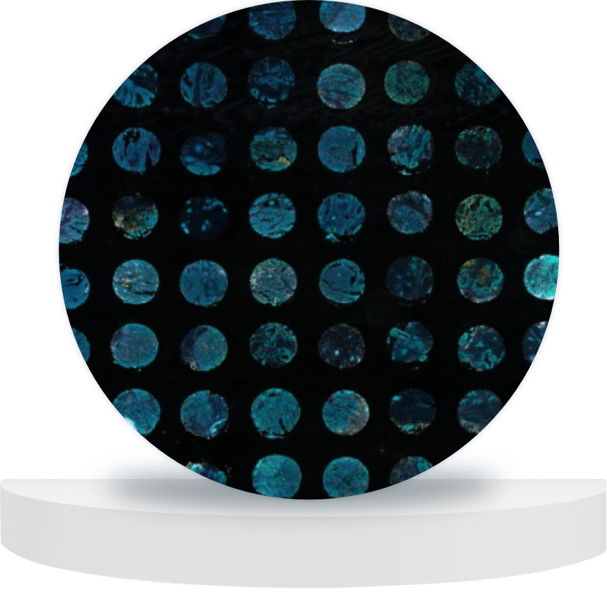
Our multiplex IHC service provides multiplexed immunofluorescent characterization of pro- and anti-tumor phenotypes and can evaluate the co-expression of multiple markers across our patient 3D tumoroids. Visualize the cell-cell and cell-extracellular matrix interactions occurring within the preserved 3D tumoroid structure.
WHY IS MULTIPLEX IHC/IF IMPORTANT?
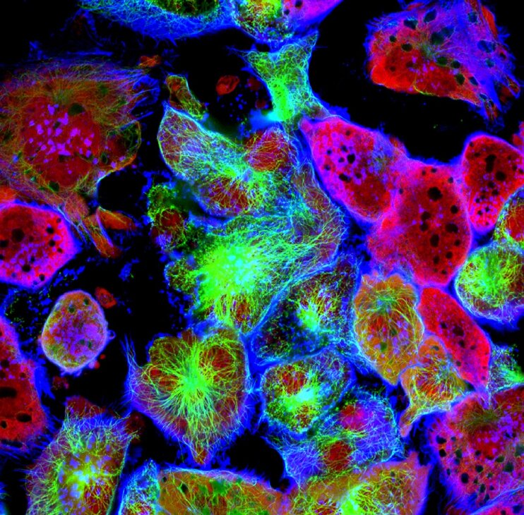
Biomarker programs need to understand the expression level and changes in functionality of key markers with drug treatment. Nilogen’s 3D-EXplore platform uses fresh patient tumor tissue derived tumoroids to enable the direct evaluation of key markers in the preserved tumor microenvironment (TME). Preserving the TME allows for the visualization of spatial interactions occurring between individual cells and with the extracellular matrix. The evaluation of a dynamic range of marker expression (e.g. PD-L1 and PD-1) in response to therapeutic intervention, allows for an in-depth investigation of the TME using our multiplex IHC service.
Capabilities
Multiplex Immunohistochemistry
Multiplex Immunohistochemistry/Immunofluorescence (mIHC/IF) provides high-throughput multiplex staining and standardized semi-quantitative analysis for highly reproducible, efficient and cost-effective tissue studies. This technique allows the simultaneous detection of multiple markers on a single tissue section, providing a comprehensive view of tissue composition, pre- and post-treatment.
Scientific Data
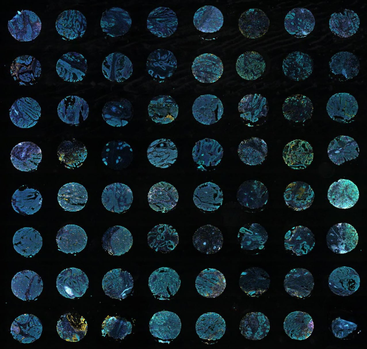
Post assay tumoroids are formalin fixed and paraffin embedded in tissue microarrays before sections are stained, imaged, and analyzed for multiple parameters.
Related Resources
Browse our latest posters and presentations using Nilogen's fresh patient tumoroid technology.
