High Content Imaging
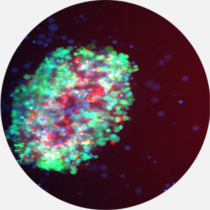
High-content assays using confocal microscopy provides phenotypic screening of living tumor tissues to simultaneously quantify markers and spatial positioning.
WHY IS HIGH CONTENT IMAGING IMPORTANT?
Combining high content imaging of multiple fluorescent tags with advanced analytics enables us to quantify penetration, tumor cell killing and phagocytosis due to drug treatment in 3D tumoroids created from fresh patient tumor tissue which retain the tumor microenvironment. These high content screening assays are unique in their ability to directly quantify the impact of advanced therapeutics in fresh tumor tissue, such as with antibody-drug conjugates delivering their toxic payload, antibody-dependent cellular cytotoxicity recruiting immune cells, CAR-T cells, as well as the penetration and replication of oncolytic virus.
Capabilities
High Content Confocal Microscopy
Quantify and match the ability of your drug to kill tumor cells with the expression of your target antigen, induce phagocytosis and measure penetration into the tumor microenvironment. Evaluate in conjunction with other proteogenomic assays to link mechanisms of action with effect.
Scientific Data
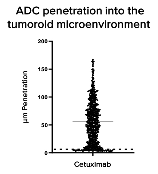

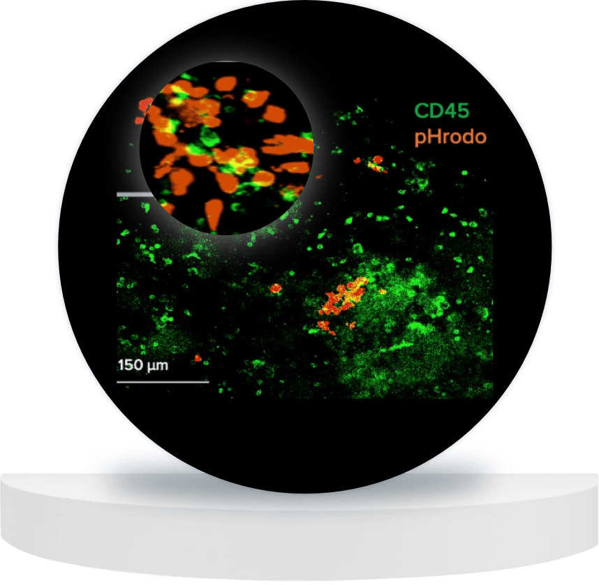
Quantifying penetration distance into the 3D tumor microenvironment of tumoroids, and ADC-mediated tumor cell killing using pHrodo-labelled cetuximab.
Related Resources
Browse our latest posters and presentations using Nilogen's fresh patient tumoroid technology.
